https://www.shroud.com/bucklin.htm
An Autopsy on the Man of the Shroud
Autopsia sobre el Hombre de la Sábana Santa - En Español
by
Robert Bucklin, M.D., J.D.
Las Vegas, Nevada
Copyright 1997
All Rights Reserved
Reprinted by Permission
For over 50 years as a Forensic Pathologist, I have been actively involved with the investigation of deaths which come under the jurisdiction of a coroner of Medical Examiner. During that time, I have personally examined over 25,000 bodies by autopsy to determine the cause and manner of death.
For most of that same period of time, I have had an abiding interest in the study of the Shroud of Turin from a medical view point. It seemed to be a natural decision for me to integrate my two interests and to try to record the results of what would have been done if the human body image on the Shroud of Turin were to be examined by a modern day Medical Examiner’s office.
The full body imprint, front and back, together with the individual characteristics of blood stains on the cloth, which represent specific types of injury, make it quite feasible for an experienced forensic pathologist to approach the examination of the Shroud image as would a medical examiner performing an autopsy on a person who has died under unnatural circumstances. It is the aim of this presentation to replicate such an autopsy examination using the image on the Shroud to delineate traumatic findings and to interpret the cause and the results of those injuries, as well as to present the most reasonable and probable cause for the death of the individual whose image is present on the Shroud of Turin.
The first step in such an examination is to document physical features of the victim as accurately as possible. In the case of the image on the Shroud, it can be stated that the deceased person is and adult male measuring 71 inches from crown to heel and weighing an estimated 175 pounds. The body structure is anatomically normal, representing a well-developed and well-nourished individual with clearly identifiable head, trunk, and extremities. The body appears to be in a state of rigor mortis which is evidenced by an overall stiffness as well as specific alterations in the appearance of the lower extremities from the posterior aspect. The imprint of the right calf is much more distinct than that of the left indicating that at the time of death the left leg was rotated in such a way that the sole of the left foot rested on the ventral surface of the right foot with resultant slight flexion of the left knee. That position was maintained after rigor mortis had developed.
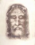
After an overall inspection and description of the body image, the pathologist continues his examination in a sequential fashion beginning with the head and progressing to the feet. He will note that the deceased had long hair, which on the posterior image appears to be fashioned into a pigtail or braid type configuration. There also is a short beard which is forked in the middle. In the frontal view, a ring of puncture tracks is noted to involve the scalp. One of these has the configuration of a letter "3". Blood has issued from these punctures into the hair and onto the skin of the forehead. The dorsal view shows that the puncture wounds extend around the occipital portion of the scalp in the manner of a crown. The direction of the blood flow, both anterior and posterior, is downward. In the midline of the forehead is a square imprint giving the appearance of an object resting on the skin. There is a distinct abrasion at the tip of the nose and the right cheek is distinctly swollen as compared with the left cheek. Both eyes appear to be closed, but on very close inspection, rounded foreign objects can be noted on the imprint in the area of the right and left eyes.
Upon examining the chest, the pathologist notes a large blood stain over the right pectoral area Close examination shows a variance in intensity of the stain consistent with the presence of two types of fluid, one comprised of blood, and the other resembling water. There is distinct evidence of a gravitational effect on this stain with the blood flowing downward and without spatter of other evidence of the projectile activity which would be expected from blood issuing from a functional arterial source. This wound has all the characteristics of a postmortem type flow of blood from a body cavity or from an organ such as the heart. At the upper plane of the wound is an ovoid skin defect which is characteristic of a penetrating track produced by a sharp puncturing instrument.
There seems to be an increase in the anteroposterior diameter of the chest due to bilateral expansion.
The abdomen is flat, and the right and left arms are crossed over the mid and lower abdomen. The genitalia cannot be identified.
By examination of the arms, forearms, wrists, and hands, the pathologist notes that the left hand overlies the right wrist On the left wrist area is a distinct puncture-type injury which has two projecting rivulets derived from a central source and separated by about a 10 degree angle. As it appears in the image, the rivulets extend in a horizontal direction. The pathologist realizes that this blood flow could not have happened with the arms in the position as he sees them during his examination, and he must reconstruct the position of the arms in such a way as to place them where they would have to be to account for gravity in the direction of the blood flow. His calculations to that effect would indicate that the arms would have to be outstretched upward at about a 65 degree angle with the horizontal. The pathologist observes that there are blood flows which extend in a direction from wrists toward elbows on the right and left forearms. These flows can be readily accounted for my the position of the arms which he has just determined.
As he examines the fingers, he notes that both the right and left hands have left imprints of only four fingers. The thumbs are not clearly obvious. This would suggest to the pathologist that there has been some damage to a nerve which would result in flexion of the thumb toward the palm.
As he examines the lower extremities, the medical examiner derives most of his information from the posterior imprint of the body. He notes that there is a reasonably clear outline of the right foot made by the sole of that foot having been covered with blood and leaving an imprint which reflects the heal as well as the toes. The left foot imprint is less clear and it is also noticeable that the left calf imprint is unclear. This supports the opinion that the left leg had been rotated and crossed over the right instep in such a way that an incomplete foot print was formed. In the center of the right foot imprint, a definite punctate defect can be noted. This puncture is consistent with an object having penetrated the structures of the feet, and from the position of the feet the conclusion would be reasonable that the same object penetrated both feet after the left foot had been placed over the right.
As the back image is examined, it becomes quite clear that there is a series of traumatic injuries which extend from the shoulder areas to the lower portion of the back, the buttocks, and the backs of the calves. These images are bifid and appear to have been made by some type of object applied as a whip, leaving dumbbell-shaped imprints in the skin from which blood has issued. The direction of the injuries is from lateral toward medial and downward suggesting that the whip was applied by someone standing behind the individual.

